High resolution Diffusion Tensor Imaging of fixed mouse hearts
In collaboration with the Institute for Experimental Cardiovascular Medicine and the Institute for Anatomy and Cell Biology we want to investigate the role of certain signaling pathways on heart development. In conditional knock out mouse models we study the structure and the function of the myocardium.
The fiber structure of the myocardium was investigated with 3D diffusion tensor imaging (DTI) in excised hearts. The hearts were fixed in formalin and embedded in agar. The spatial resolution of the DTI sequence was 0.16 mm x 0.17 mm x 0.47 mm. Scan time was 13 h. Data processing was performed with the DTI&Fibertracking-toolbox developed in-house by the Postprocessing research group with modifications made by AMIR. The Global Fibertracking algorithm was used (Reisert et al., 2011).

MR fiber tracking image of a mouse heart
MR fiber tracking image (color-coded by orientation: red left-right, green back-front, blue top-bottom) of a formalin-fixed mouse heart showing the myocard fibers of left and right ventricle (lv, rv). Images were created with nora (www.nora-imaging.com), a web-based medical image analysis platform developed by the Postprocessing group.
Ref.: M.Reisert, I. Mader, C. Anastosoulus; M. Weigel, S. Schnell; V.Kiselev, "Global Fiber Reconstruction Becomes Practical" Neuroimage. 2011 Jan 15;54(2):955-62.
DTI fiber data of mouse heart with overlaid fractional anisotropy map
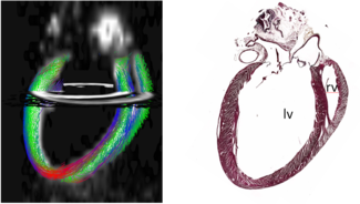
Left: Slice (0.5 mm) of the DTI fiber data in a fixed mouse heart (color-coded by orientation) in the long axis view. The fiber data is overlaid on a mask based on the fractional anisotropy map (white). Images were created with nora (www.nora-imaging.com), a web-based medical image analysis platform developed by the Postprocessing group. Right: Corresponding histological slice (HE staining). lv, rv= left/right ventricle

Color coded maps of the fractional anisotropy (FA) in formalin-fixed hearts of a conditional knock out (k.o., left) and a control mouse (right). The maps show lower (i.e. less restricted diffusion) and less homogen FA values in the myocardium in k.o. heart vs. control.
Responsible scientists:
- Dr. Claudia Weidensteiner
- apl. Prof. Dr. Dominik von Elverfeldt
- Thomas Bienert
- Dr. Jochen Leupold
Internal cooperation:
- Dr. Marco Reisert
- Institute for Experimental Cardiovascular Medicine
- Institute for Anatomy and Cell Biology

apl. Prof. Dr. Dominik von Elverfeldt
Head of AMIR
Tel.: +49 761 270-38320
E-Mail: dominik.elverfeldt@uniklinik-freiburg.de
University Medical Center Freiburg
Dept. of Radiology · Medical Physics
Killianstr. 5a
79106 Freiburg