MR Microscopy
The current method of choice for highly-resolved morphological characterization of the human tissue remains histology. If spatial resolution, contrast and sensitivity are sufficient, MR microscopy can be used as a powerful instrument for in vivo investigation as an alternative to invasive histopathological sampling.

Figure 1: Left: Representative axial MR image through skin biopsy revealing the epidermis and dermis (7 Tesla + Mousehead two-element quadrature cryo coil, res. 30×30×120 µm3, Tscan = 19 min 48 s); Right: Corresponding immunohistochemical investigation (Hematoxylin Eosin staining) of a similar skin sample validating the MRI results.

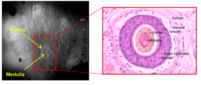
Figure 2: Left: Coronal MR image depicting the skin structure (9.4 Tesla + phased-array microcoil (IMTEK), res. 40×40×300 µm3, Tscan = 8 min 12 s); Right: Histological view of a hair follicle with its hair shaft and the surrounding epithelial sheath. (http://www.vetmed.vt.edu/education/curriculum/vm8054/labs/lab15/lab15.htm)

Figure 3: Visualization of neuronal structures in a fixed Organotypic hippocampal slice culture (OHSC) using MR microscopy in comparison to optical microscopy of histologically stained cryosections. Left: MR Image (7 Tesla + Mousehead two-element quadrature cryo coil, res. 16×18×150 µm³, Tscan = 2 h 8 min) of a fixed OHSC,; Middle: Immunohistochemical staining of 20 µm thick OHSC cryosections; Right: Nissl-stained cryosection of 20 µm thick OHSC.
Responsible scientists:
- Jochen Leupold
- Former member: Katharina Göbel
- Former member: F. Kording
Internal cooperation:
Further cooperation:
apl. Prof. Dr. Dominik von Elverfeldt
Head of AMIR
Tel.: +49 761 270-38320
E-Mail: dominik.elverfeldt@uniklinik-freiburg.de
University Medical Center Freiburg
Dept. of Radiology · Medical Physics
Killianstr. 5a
79106 Freiburg


