MR imaging of Vasculature Biomarkers in Glioma
State-of-the-Art:
- combined imaging of vessel size imaging (VSI) and DCE-MRI biomarkers in a mouse model
- study with mouse tumor model to characterize the changes in neovasulature under therapy in preparation
New Developments:
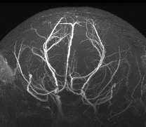
- TOF-Angiography in a mouse featuring a high in-plane resolution (50µm)
- postprocessing: Segmentation of the tumor to account for GBM heterogeneity

Figure 1: Exemplary T2-weighted RARE image (a) and corresponding segmentation (b), VSI (c) and Ktrans map (d)
Responsible scientists:
- Wilfried Reichardt
- Jochen Leupold
- Former member: F. Kording
Internal Cooperation:
Further cooperation:

Dr. med. Wilfried Reichardt
Contact person
Tel. +49 761 270-73910
E-Mail: wilfried.reichardt@uniklinik-freiburg.de
University Medical Center Freiburg
Dept. of Radiology · Medical Physics
Killianstr. 5a
79106 Freiburg