Neuro navigation - further details
Neuro navigation allows the exact localisation of areas on or in the patien’s body – similar to GPS navigation in traffic. For this purpose, precise image data of the body are necessary, for example MRT (magnetic resonance tomography) or CT (computer tomography).
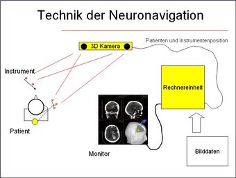
Those pictures are loaded into a computer program. The patient’s position is located with a 3 D camera in the room. The position of the patient and the surgical instruments are made visible through infrared diodes. therefore, it is possible to visualize on the screen the exact position of the instrument within the image data of the patient. So the neurosurgeon can see what kind of structures are there in the surrounding area and where exactly the tumour is located.
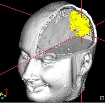
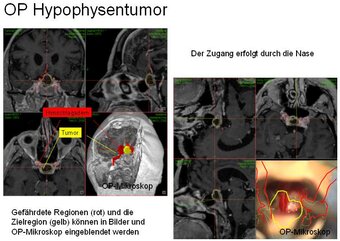
Before surgery, the area of interest is determined. This can be an abnormal area of the brain, a tumour, or a vascular malformation. In addition, highly important and endangered regions can be marked. So they are easier to recognise during surgery. Furthermore, they can be shown in the operation microscope which is linked to the navigation system.
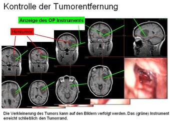
With the help of this technique, the surgery can be performed safer. Through picturing the current position of the operating instruments, one can see what lies hidden under the surface. The reduction of a tumour, for example, can be followed until the very edge.
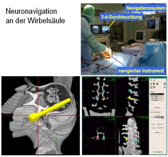
In case of difficult anatomic situations, neuro navigation is used also for spine procedures. During surgery, image data similar to a CT are produced with a 3 D X-ray fluoroscopic equipment. These image data are transferred to the navigation system. The employed instruments and the patient’s position are followed by the 3 D camera of the navigation system. Thus, the implantation of screws, for example, can be planned and placed with millimetre precision.

Prof. Dr. Ulrich Hubbe, MD
Senior neurosurgeon

Prof. Dr. Jan-Helge Klingler, MD
Senior neurosurgeon
Consultation hours for spine surgery


