Innate immunity group
Head: M.PrinzOur group focuses on the role of the brain specific innate immune system. Important molecules involved in innate immunity are chemokine receptors, Toll-like receptors (TLRs) and cytokines such as interferons. Major player of this system within the brain are brain macrophages (microglia) which serve as the first barrier for invading pathogens. We are currently investigating the mechanisms by which microglia contribute to the induction and resolution of brain damage using mouse models of multiple sclerosis (EAE, cuprizone model) and neurodegeneration, e.g. Alzheimers disease.
Personalia
| Prof. Dr. Marco Prinz | Principal Investigator | +49-761-270-51060 |
| Dr. Lukas Amann | Postdoc | +49-761-270-63114 |
| Dr. Chintan Mukeshbhai Chhatbar | Postdoc | +49-761-270-93380 |
| Dr. James Cook | Postdoc | +49-761-270-63111 |
| Dr. Anaelle Dumas | Postdoc | +49-761-270-63112 |
| Dr. Elena Guffart | Postdoc | +49-761-270-63091 |
| Dr. Michael Schulz | Postdoc | +49-761-270-63137 |
| Dr. Han Gao | Postdoc | +49-761-270-63136 |
| Maximilian Fliegauf | Phd Student | +49-761-270-63113 |
| Adrià Dalmau Gasull | Phd Student | +49-761-270-63112 |
| Nishita S. Ghariwala | Master Student | +49-761-270-63112 |
| Takashi Shimizu | Guest Researcher | +49-761-270-63136 |
| Saskia Wundt | Technician | +49-761-270-63114 |
| Maria Oberle | Technician | +49-761-270-50740 |
FUNCTION OF MICROGLIA IN CNS INFLAMMATION
WHAT IS THE ROLE OF THE INNATE IMMUNE SYSTEM INSIDE THE BRAIN?
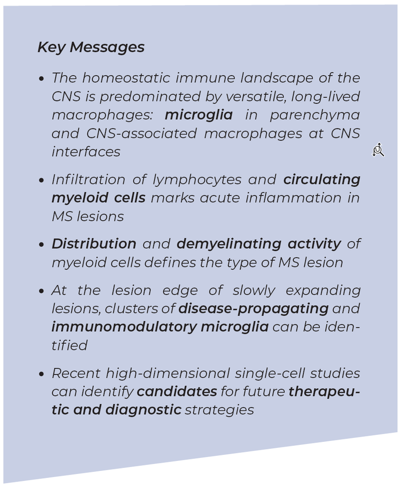
We know that macrophages are present throughout the entire body and play an important role in the adaptive immune response. Inside the brain parenchyma (i.e. functional tissue comprising neurons and glia cells), innate immunity is mediated by specialized long-lived tissue macrophages, called microglia. Microglial cells can live for several years, are highly active and extremely motile. On one hand, microglia act as the „watchdogs“ of the brain, looking for potential pathogens and protecting the brain. On the other hand however, microglia can play disease-inducing roles, promoting brain inflammation and neurodegeneration. Indeed, several dozen microglia-linked risk alleles for CNS diseases have already been identified, implicating microglia for example in the pathophysiology of Alzheimer’s disease, Parkinson‘s disease and multiple sclerosis (MS). In addition to microglia, other types of macrophages are also present in the outer boundaries of the brain, residing in the leptomeninges, the perivascular space, and the choroid plexus. Normally, in a healthy brain, a balance (i.e. homeostasis) between these cell types prevails. When an inflammation occurs, it evokes a CNS autoimmune response and this balance is lost. During inflammation, the number of macrophages expands and crucially, circulating cells from the blood may also enter the brain via a disrupted blood-brain barrier, leading to a mixture of various cell types within the brain. For example, in MS there is a sustained neuroinflammation that is characterized by the presence of blood-derived macrophages, B-cells and T-cells.

WHAT IS THE ROLE OF THE INNATE IMMUNE SYSTEM IN MS?
Characteristic histological features of MS lesions were first identified over 100 years ago, reporting the presence of small round infiltrating cells and the absence of white matter. More modern histological classifications now rely on presence and distribution of bloodderived macrophages and microglia to determine the activity degree of the lesion (i.e. active, mixed active/inactive, inactive, each with or without ongoing demyelination). Active, demyelinating lesions are characterized by the presence of microglia and bloodderived macrophages throughout the lesion. Neuroimaging also shows that iron is accumulated within microglia and blood-derived macrophages located at MS lesions, forming iron-rims. Iron liberation and its accumulation are likely to occur during demyelination, primarily in lesion types identified as “slowly expanding lesions” and “paramagnetic rim lesions”.


NEW INSIGHTS FROM SINGLE-CELL ANALYSIS
Single-cell analysis is a method that can be used to better understand disease pathogenesis, allowing to analyze individual cells at protein- or RNA-level, as opposed to classic bulk analyses where cells are analyzed and averaged together. Single-cell analysis is mostly employed in cancerous diseases, especially for tumors outside of the brain, but has recently become available for cells within the brain (as investigated in the laboratory of Prof. Prinz). For example, single-cell analysis can inform in more detail on the composition of brain lesions, identifying specific clusters for various cell-types (e.g. subsets of disease-associated microglia, lymphocytes, macrophages).
Single-cell studies carried out in MS patients demonstrated the heterogeneity of microglial cells in MS lesions, describing microglia clusters with different activation and immunoregulatory patterns. Such studies identified microglial cells as the most activated cell type in chronic active lesions (specifically in paramagnetic rim lesions) and identified the complement factor 1q as an important mediator of microglia activation.
These kinds of data are very descriptive, but it is important to have a better picture of the disease in order to have a better therapy one day.
«Our hope is that this technology will identify potential candidates for future therapeutic approaches targeting specific cell states that are only found during new inflammation.»
Publications
1. Masuda T, Amann L, Monaco G, Sankowski R, Staszewski O, Krueger M, Del Gaudio F, He L, Paterson N, Nent E, Fernández-Klett F, Yamasaki A, Frosch M, Fliegauf M, Bosch LFP, Ulupinar H, Hagemeyer N,Schreiner D, Dorrier C, Tsuda M, Grothe C, Joutel A, Daneman R, Betsholtz C, Lendahl U, Knobeloch KP, Lämmermann T, Priller J, Kierdorf K, Prinz M (2022) Specification of CNS macrophage subsets occurs postnatally in defined niches. Nature, 604, 7907, 740-748
2. Masuda T, Amann L, Sankowski R, Staszewski O, Lenz M, D Errico P, Snaidero N, Costa Jordão MJ, Böttcher C, Kierdorf K, Jung S, Priller J, Misgeld T, Vlachos A, Meyer-Luehmann M, Knobeloch KP, Prinz M (2020) Novel Hexb-based tools for studying microglia in the CNS. Nat Immunol, 21, 7, 802-815
3. Masuda T, Sankowski R, Staszewski O, Böttcher C, Amann L, Sagar, Scheiwe C, Nessler S, Kunz P, van Loo G, Coenen VA, Reinacher PC, Michel A, Sure U, Gold R, Grün D, Priller J, Stadelmann C, Prinz M (2019) Spatial and temporal heterogeneity of mouse and human microglia at single-cell resolution. Nature, 566, 7744, 388-392
4. Sankowski R, Böttcher C, Masuda T, Geirsdottir L, Sagar, Sindram E, Seredenina T, Muhs A, Scheiwe C, Shah MJ, Heiland DH, Schnell O, Grün D, Priller J, Prinz M (2019) Mapping microglia states in the human brain through the integration of high-dimensional techniques. Nat Neurosci, 22, 12, 2098-2110
5. Geirsdottir L, David E, Keren-Shaul H, Weiner A, Bohlen SC, Neuber J, Balic A, Giladi A, Sheban F, Dutertre CA, Pfeifle C, Peri F, Raffo-Romero A, Vizioli J, Matiasek K, Scheiwe C, Meckel S, Mätz-Rensing K, van der Meer F, Thormodsson FR, Stadelmann C, Zilkha N, Kimchi T, Ginhoux F, Ulitsky I, Erny D, Amit I, Prinz M (2019) Cross-Species Single-Cell Analysis Reveals Divergence of the Primate Microglia Program. Cell, 179, 7, 1609-1622 e1616
6. Jordão MJC, Sankowski R, Brendecke SM, Sagar, Locatelli G, Tai YH, Tay TL, Schramm E, Armbruster S, Hagemeyer N, Groß O, Mai D, Çiçek Ö, Falk T, Kerschensteiner M, Grün D, Prinz M (2019) Single-cell profiling identifies myeloid cell subsets with distinct fates during neuroinflammation. Science, 363, 6425eaat7554
7. Tay TL, Mai D, Dautzenberg J, Fernández-Klett F, Lin G, Sagar, Datta M, Drougard A, Stempfl T, Ardura-Fabregat A, Staszewski O, Margineanu A, Sporbert A, Steinmetz LM, Pospisilik JA, Jung S, Priller J, Grün D, Ronneberger O, Prinz M (2017) A new fate mapping system reveals context-dependent random or clonal expansion of microglia. Nat Neurosci, 20, 6, 793-803
8. Goldmann T, Wieghofer P, Jordão MJ, Prutek F, Hagemeyer N, Frenzel K, Amann L, Staszewski O, Kierdorf K, Krueger M, Locatelli G, Hochgerner H, Zeiser R, Epelman S, Geissmann F, Priller J, Rossi FM, Bechmann I, Kerschensteiner M, Linnarsson S, Jung S, Prinz M (2016) Origin, fate and dynamics of macrophages at central nervous system interfaces. Nat Immunol, 17, 7, 797-805
9. Erny D, Hrabě de Angelis AL, Jaitin D, Wieghofer P, Staszewski O, David E, Keren-Shaul H, Mahlakoiv T, Jakobshagen K, Buch T, Schwierzeck V, Utermöhlen O, Chun E, Garrett WS, McCoy KD, Diefenbach A, Staehel P, Stecher B, Amit I, Prinz M (2015) Host microbiota constantly control maturation and function of microglia in the CNS. Nat Neurosci, 18, 7, 965-977
10. Kierdorf K, Erny D, Goldmann T, Sander V, Schulz C, Perdiguero EG, Wieghofer P, Heinrich A, Riemke P, Hölscher C, Müller DN, Luckow B, Brocker T, Debowski K, Fritz G, Opdenakker G, Diefenbach A, Biber K, Heikenwalder M, Geissmann F, Rosenbauer F, Prinz M (2013) Microglia emerge from erythromyeloid precursors via Pu.1- and Irf8-dependent pathways. Nat Neurosci, 16, 3, 273-80
